Stories

- Article
Two health centres, two ideologies
Two futuristic, light-filled buildings aimed to bring forward-looking healthcare to city dwellers. But the principles behind each were very different.

- Article
Conserving Audrey
Elena describes how specially designed storage allows Audrey’s scrapbooks to retain all traces of her creative process, although their intrinsic fragility means deterioration is almost inevitable.
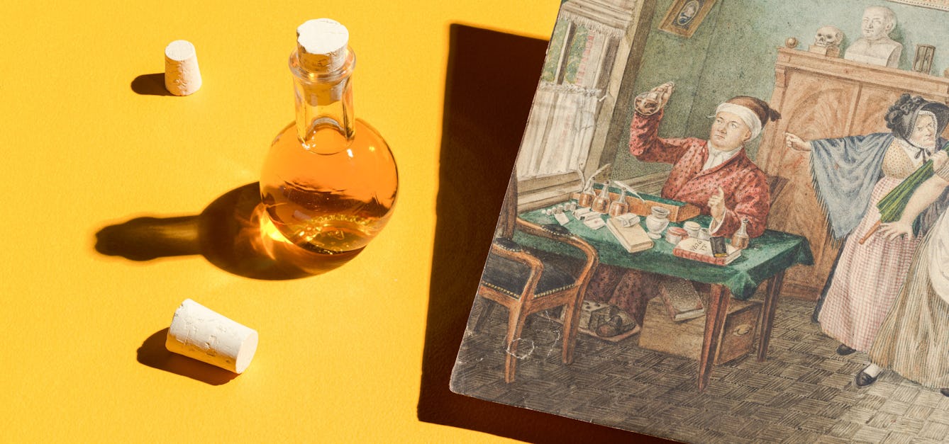
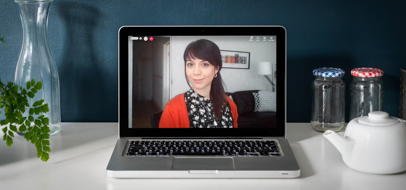
- Article
Remote diagnosis from wee to the Web
Medical practice might have moved on from when patients posted flasks of their urine for doctors to taste, but telehealth today keeps up the tradition of remote diagnosis – to our possible detriment.
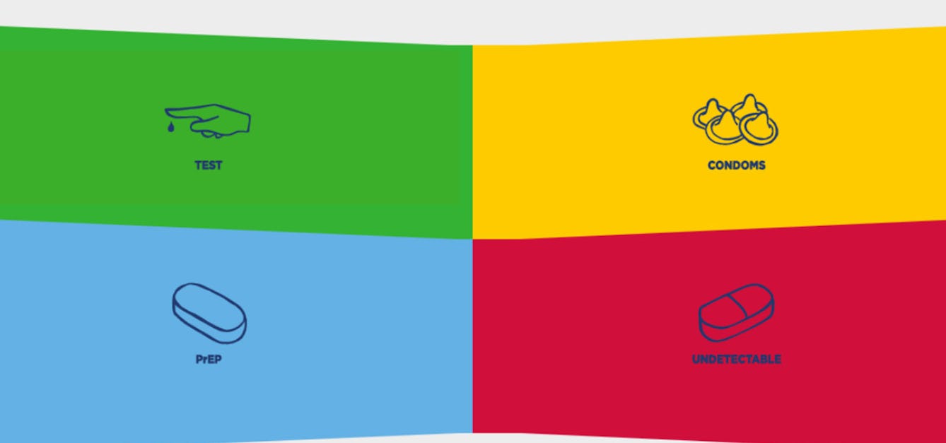
- Interview
How to design an HIV awareness campaign
Using carefully crafted, colourful graphics is one public health team’s creative approach.
![Clocks: an elaborately-cased mantel clock, with the figure of a dairymaid, and various bases (above). Coloured lithograph, [c.1875?].](https://iiif.wellcomecollection.org/image/V0024692/full/282%2C/0/default.jpg)


Catalogue
- Archives and manuscripts
Bases para el establecimiento
Date: c.1869Reference: WMS/Amer.121/20Part of: Mexico: Casa de Maternidad- Books
Bases neurobiologiques des réflexothérapies / Jean Bossy.
Bossy, JeanDate: 1978- Books
Bases farmacológicas de la terapéutica medicamentosa / F.G. Valdecasas [and others].
Date: 1969- Books
Bases para la organización de los servicios sanitarios / por Adolfo Serigó Segarra.
Serigó Segarra, Adolfo.Date: 1972- Books
Bases históricas de la psiquiatría catalana moderna / Edelmira Domenech, Jacint Corbella, Dídac Parellada (editors).
Date: 1987